What is the 12 week NT scan. Your 12-week ultrasound is one of your most important prenatal appointments At 12 weeks your baby has organs and tissue that are starting to grow rapidly.

All About Normal 12 Week Ultrasound Ultrasoundfeminsider
The Nub Theory is a popular way for parents-to-be to predict their babys gender at just 12 weeks.

Ultrasound at 12 weeks. Of course accuracy is a loose term but at least youll have a fairly good idea Check-in with how the baby growing process is going. 12 Weeks Pregnant Ultrasound. Reasons for an ultrasound during week 12 of pregnancy.
How well is conception date accuracy with ultrasound at 12 weeks. To test the possibilities of congenital chromosomal abnormalities. You can expect to see your baby moving at this ultrasound which is exciting.
If u have an ultrasound at 12 weeks and it comes back saying u are 12 weeks 4 days - is that enough of a difference to move up your due date Answered by Dr. If CRL 50 mm gestational age 114 the gender cannot be reliably predicted. By the end of the third month there are many reasons to have an ultrasound but the most common and major reasons are.
For many women an ultrasound or dating scan around twelve weeks provides the first. The twelfth week of pregnancy is an important and exciting milestone. A general check of the mother and foetuss internal organs and structures.
One can get the ultrasound done anytime between the 11th and 13th week. The 12 week ultrasound is also called the dating scan. Has anybody went to their 12 week appointment and not had an ultrasound.
At three months gestational age doctors typically have a clear view of the fetus in the womb. Week 12 Ultrasound. I had one at 7 weeks but thats it.
It is difficult to calculate pregnancy age yourself. Unlike other methods that use astrology to predict a babys gender or old wives tales about pregnancy to try and guess if you are carrying a boy or a girl the Nub Theory uses the 12-week ultrasound scan picture to predict the sex of your baby more accurately. My lmp started on nov28 2011 lasting five days.
Though often referred to as the twelve week scan it may happen between eight and 14 weeks. To check whether the foetus is at a risk of developing physical deformities. It marks the end of the first trimester one down two to go and at this point your baby is fully formed right down to the teeny-tiny toes.
During the examination the fetus is seen by abdominal ultrasound. Male gender may already be reliably determined when CRL 55 mm gestational age 120. And my next appointment isnt until Im 16 weeks.
The foetuss length specifically from its head to its bottom. I had an ultrasound on jan17 2012 counting me at 7 weeks and 1 day then another ultrasound on feb21 2012counting me at 12 weeks exa Answered by Dr. During the 12-week ultrasound which takes place between weeks 11 and 13 of your pregnancy theyll be snapping sonogram images to review and will be looking for several things including.
This is known as a Crown Rump length. Your 12-week scan can take place any time between 11 and 13 weeks. They can also open and close their fingers.
The 12 week scan is a routine ultrasound examination carried out at 10 to 14 weeks of gestation. Its amazing to think that at this point your baby is. At 12 weeks pregnant you will be offered an ultrasound scan.
Their brain is also getting larger and more developed by the day. Your due date can be pretty accurately predicted at your 12 week ultrasound. Occasionally the view is not clear and it may be necessary to perform a vaginal scan.
The ultrasound dating now is remarkably accurate. Ultrasound evaluation offetal gender at 12-14 weeksFetal gender may reliably be determined when CRL 60 mm gestational age 122. Due date is 2 weeks on either side of the estimated date.
What It Would Look Like Your baby is a full three inches long this week. Your doctor will schedule and let you know the exact date to come for 12 weeks ultrasound scan. What else is measured during a 12 week ultrasound.
Find out more about the size and all the other exciting developments going. Was so hoping I would get to see baby today to ease my worries but they didnt do an ultrasound or even try to listen to the heart beat.
3D4DHD Ultrasound at 37 weeks- good or bad idea. UC Baby 3D Ultrasound from 18 week to 37 week of pregnancy - YouTube.

3d 4d Hd Ultrasound At 37 Weeks Good Or Bad Idea May 2017 Babies Forums What To Expect
But what they.

3d ultrasound 37 weeks. About Press Copyright Contact us Creators Advertise Developers Terms Privacy Policy Safety How YouTube works Test new features Press Copyright Contact us Creators. I am getting a 3D ultrasound done and I am 37 weeks. Your baby will be much bigger and an ultrasound is done in order to give the following information.
What It Would Look Like Now that youre 37 weeks pregnant your baby is full term. Week 37 Ultrasound. Now Im worried and plan to bring pic to next doctors visit on Monday to ask if it looks ok.
I wasnt told about having a full bladder so would I still have to have a glass of water a half hour before my appointment like my 20 week ultrasound or do I not need to have a full bladder being farther along. BABY IS ALMOST HERE YALL 3 more weeks Also THANK YOU SO MUCH FOR 152K you guys are truly the bestOu. I made the mistake of spending 200 on a 3d-4d ultrasound.
After all youre going to have a baby soon and your body knows it so its getting ready. Has anyone gotten one done this late in the game before. 3D ultrasound at 37-38 weeks.
Vor 1 Jahrzehnt. My doctors office doesnt do another unless its medically indicated they did however already check to see if babys head is down- and it is but thats all they looked at and part of me. Fulton has grown about 1lb since the 35 week ultrasound.
Full bradder is not necessary. Yawns stretches and even smiles are often captured in our 3D4D 5D ultrasound sessions. We also received some encouraging news.
View the unbelievable clarity of this 4d Video clip of an ultrasound session taken at 36 weeks and 5 days at GoldenView Ultrasound. At this stage of pregnancy most women will have had all their ultrasounds already. Has anyone gotten one done this late in the game before.
Hey loves welcome or welcome back to my channel. The fetuss presentation - wether the babys head is down breech or up. At 37 weeks youre full term and nearly at the end of your pregnancy.
I would think that the doctors office would be notified right away if there was an issue and they would get in touch with. Holding out hope for a miracle. At this stage the baby has put on some weight and filled out to make features more visible yet still enough fluid in front of babys face to obtain great.
However we do recommend a gestational age of 26-34 weeks for the best facial detail. At 37 weeks mild contractions are not out of the ordinary. He currently weights about 315lbs up from 214 just 2 weeks ago.
Hi all So my last ultrasound was the one done at 20weeks- the anatomy scan and gender reveal and Im wondering if its worth getting a 4D one done at 37 weeks. Thank You Lord for such blessings. My doctors office doesnt do another unless its medically indicated they did however already check to see if.
I had an ultrasound at 29 weeks and I noticed when I was home that the back of his head looks all pointy not 3D normal ultrasound which was not the case on other ultrasounds. At Elises 37 week ultrasound we were able to capture some beautiful imagines of baby Fulton Josephs face. Due to anticipation for labor your baby has probably dropped by now into the pelvic area which can cause an increased in abdominal and pelvic pressure.
3D 5D ultrasound images and 4D ultrasound video can be obtained at any stage. 3d 4d ultrasound at 36 weeks GoldenView Ultrasound - YouTube. 37 weeks pregnant ultrasound Your ultrasound schedule will vary based on where you live how your pregnancy is going and who your doctor is.
While it was amazing to see my son the tech was very inexpirenced and nobody told me at 35 weeks. Ultrasound at 37 weeks. This is our first baby and shes beautiful.
Normal pregnancy symptoms at 37 weeks. So my last ultrasound was the one done at 20weeks- the anatomy scan and gender reveal and Im wondering if its worth getting a 4D one done at 37 weeks. Learn whats going on with your little one at this part of your journey together.
Watch the change of baby from 18 week tp 37 weeks of pregnancy on this special video created by UC Babys 3D ultrasound. This is the most significant growth he has demonstrated in at least 6 weeks.
But in medical gynecologic practice cases when additional research of the fetus on indications is required very often meet. In some countries sonograms are performed just twice during pregnancy.
/babyboyultrasound-7bf2ced4b4794754b67dea974b7ec744.jpg)
What To Look For In Your Baby Boy Ultrasound
It is in fact the preparing phase of the baby to take up the challenge of the new world where it is going to be born.
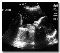
34 week pregnancy ultrasound. What It Would Look Like Now that baby is at 34 weeks she can now see hear learn and remember. See how your baby is developing at 34 weeks of pregnancy. Once at 16 - 18 weeks to assess fetal abnormalities and again at 32 - 34 weeks to assess age and well-being.
At 34 weeks of pregnancy the woman enters a new phase of pregnancy which is often recognized by descending of the twins towards the pelvis. The special nonstress test is done to measure the babys heart rate over a period of time. If hes born this week.
Week 34 Ultrasound. Find out all about these important pregnancy milestones. Piling on the fat.
Your first prenatal ultrasound which is scheduled sometime between the 6th and 9th week confirms your pregnancy determines due date and monitors the heartbeat. 1 This week baby weighs about 5 pounds 2312 grams. With the help of ultrasound images your gynaecologist would take several decisions regarding the birthing process.
It is now recommended that all pregnant women have a dating scan in the first trimester - ideally at 10 to 13 weeks of pregnancy - to confirm your dates. When the babys head turns towards the mothers vaginal canal the upper portion of uterus reduces in size. The ultrasound tech said approx 6 pounds for my baby boy and in the 80th percentile.
Baby development at 34 weeks. Your babys fat layers which will help regulate his body temperature once hes born are filling him out making him rounder. 34 weeks pregnant ultrasound.
At pregnancy week 34 the baby is primarily focused at growing and hence spends most of its time in filling out and adding meat to its bones. Also if you recently had a babes and know what they measured at 34 weeks. This is a combination of an ultrasound done at 34 weeks pregnant and a special nonstress test.
She weighs more than 21kg 47lb and is about 45cm 177in from head crown to heel. Besides that there should only be two ultrasounds. Ultrasound at 34 Weeks Pregnant Planned ultrasonic researches by 34th week of pregnancy are as a rule already passable.
34 Weeks Pregnant Ultrasound Baby Movement Weight. If your doctor orders it you could have a biophysical profile BPP which is a combination of a 34 weeks pregnant ultrasound and a special non-stress test which measures babys heart rate over a period of 20 minutes. How much did they end up coming out at.
No unless towards the end they believe baby is breech and need an ultrasound to confirm. At 34 weeks a baby is over 12 14 inches 311 centimeters from the top of their head to the bottom of their buttocks known as the crown-rump length and babys height is nearly 17 12 inches 442 centimeters from the top of their head to their heel crown-heel length. TwinMultiple Pregnancy Ultrasound at 34 Weeks An ultrasound is conducted to check the health of the babies and ensure safe delivery.
Your baby is the size of a cantaloupe melon. During pregnancy week 34 the baby is around 177 inches in length and usually weighs in at around 47 pounds. Considering that nearly 1 of births every year are known to have congenital heart defects the doctor observes the structure of the heart to check for any congenital disabilities in the second ultrasound or the 20th-week.
At your 34 week ultrasound how much was baby measuring and what percentile. 34 Weeks Pregnant Ultrasound Youll likely take a trip to the OB this week since youre probably seeing them every other week. In this stage the mother who is carrying the baby feels some.
If your doctor orders it you may do a biophysical profile BPP. The one at around 12 weeks and another at 20. In this phase of pregnancy in 34th week the baby inside the womb of the regnant woman gets gradually stronger.
I found out at 19wks on a regular Ultrasound machine you can find out as early as 15wks but they recommend at least 18wksMy guess is at 18wks on a. 18 Weeks Ultrasound Twins Carrying Out Analyses Generally they carry out the analysis of urine to learn about work of kidneys and blood test is hemoglobin level in.
18 Weeks 3d 4d Hd Live Ultrasound
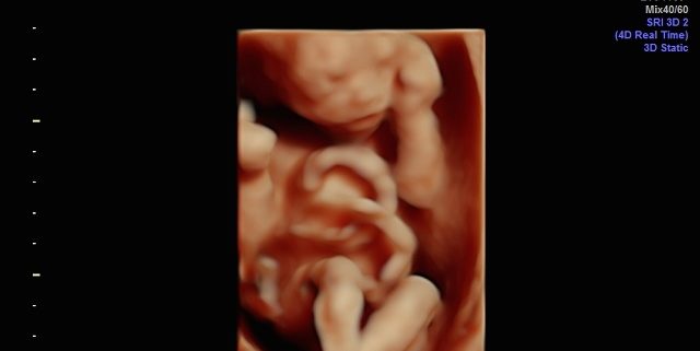
What is a 4D ultrasound.

18 week 4d ultrasound. This ultrasound is often called an anatomy scan. What Is A 4d Ultrasound Test. A 22-week ultrasound scan can help monitor the health of the fetus and the pregnancy and 3D and 4D ultrasounds can provide more detail.
A 4D ultrasound takes this experience to the next level. If you dont get to peek inside the womb this week youll probably get the chance within the next few weeks. 3-D and 4-D ultrasounds can seem like the perfect solution to get a sneak peek of your little one.
As a soon-to-be mother youre probably very excited to see your baby every time you feel them kick inside your stomachYou can do that by getting an ultrasound which can be 2D 3D Doppler and 4D depending on how much detail you want to see. With a transabdominal exam the sonographer. Follow me on Instagra.
At 18 weeks your baby has mastered the art of yawning along with hiccuping which you may feel soon. Between weeks 18 and 20 a trained sonographer will perform a detailed anatomy scan called a level 2 ultrasound. Snuggling cord at 18 weeks.
It is possible you may be scheduled for a more comprehensive and detailed ultrasound. A wide variety of 18 week 4d ultrasound options are available to you such as quality certification material and shelf life. The 4D ultrasound uses sound waves to create this moving image.
It is recommended that you have an ultrasound between 18 to 20 weeks. This is my first baby. Cannot tell the gender yet coz the baby is not cooperating during ultrasound need to resched.
The 18th week of pregnancy is usually the earliest that a healthcare provider can do the anatomy scan. You may catch a glimpse of that adorable yawn and all those other fetal movements at your ultrasound. In a 3D ultrasound you can see a three-dimensional image of your baby.
18 to 20 Week Ultrasound. It is at this time that the sonographer will measure the size of your baby check the major organs measure the level of amniotic fluid to make sure that its. This week you may feel some of those moves and get to see your child demonstrate them on the ultrasound screen.
At 18 weeks your baby is growing and moving all around. Ultrasound 4D HD live performed in the Ultrasound Unit of the Gynecological Clinic of Dr. 3D 5D ultrasound images and 4D ultrasound video can be obtained at any stage.
A 4D ultrasound test 1 is a way of reproducing a moving image of your baby inside your womb. Rafael Ortega Muñoz in which the face of a fetus of 18 weeks in 4D. Forty weeks can feel like a lifetime to wait to see your baby.
At 18 weeks your baby-to-be is large enough that your sonographer will perform the ultrasound transabdominally. The purpose of this ultrasound is to be sure that your fetus is developing normally. On the next dayOUR PRECIOUS.
At this stage the baby has put on some weight and filled out to make features more visible yet still enough fluid in front of babys face to obtain great images. During the 18-week ultrasound a doctor or ultrasound technician will use an ultrasound. 18 week HD Live4D ultrasound.
Upon seeing one of these ultrasounds Ive heard parents say Oh hes got his grandfathers nose or She looks just like her sister when she was born. However we do recommend a gestational age of 26-34 weeks for the best facial detail. 18w4d Detailed ScanThis video is 3 individual clips of our little angel in 4D.
I have also attached a few 2d ultrasound pics as well.
They often have leakage across the tricuspid valve and reverse flow in the ductus venosus. Dilatation of the kidneys pyelectasis.

A Review On Techniques For Computer Aided Diagnosis Of Soft Markers For Detection Of Down Syndrome In Ultrasound Fetal Images Biomedical And Pharmacology Journal
Down Syndrome can include cardiovascular central nervous craniofacial musculoskeletal gastrointestinal and urinary tract system anomalies.

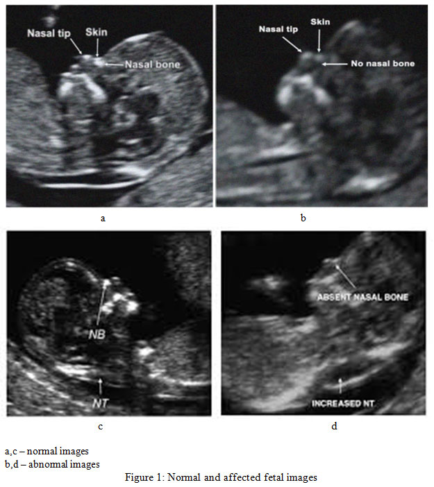
What does a down syndrome ultrasound look like. What are some of the signs and ultrasound findings of down syndrome. Thickened nuchal fold nuchal translucency Duodenal Atresia double bubble Echogenic bowel. The following are ultrasound markers that are seen more frequently in fetuses with Down syndrome.
An ultrasound technician will gently move a. For most people with Down syndrome this anomaly causes a host of distinctive physical characteristics as well as potential health and medical problems. Also the lining would be thin without any masses.
Ultrasonography is the imaging modality mainstay of prenatal screening and diagnosis of Down syndrome and it is often used in combination with biochemical tests. Certain second trimester markers for Downs syndrome that are detected during an ultrasound exam are more meaningful than other markers. Ultrasound is a key component of aneuploidy screening.
Babies with Downs syndrome are more likely to have a small or absent nose bone with a flat profile. Prenatal ultrasound findings were reviewed in 94 consecutive fetuses with proved Down syndrome trisomy 21 during a 6-year period at a single institution. The most predictive finding of Down syndrome on prenatal ultrasound is an absent nasal bone.
This ultrasound uses high frequency sound waves that generate a moving picture of your internal workings and your babys changing form. The nasal bone can be a good marker for Downs syndrome agrees Rosemary Reiss who practises maternal fetal medicine at Brigham and Womens Hospital in Boston Massachusetts. What does a down syndrome ultrasound look like.
The FP line was defined as the line that passes through the midpoint of the anterior border of. One or more abnormalities were found in 31 fetuses 33 including two of 11 fetuses seen before 14 weeks 17 of 68 fetuses seen between 14-24. A Verified Doctor answered.
Your age the level of two hormones free ß-hCG and PAPP-A in your blood and. The entire uterus would look the same with out any fibroid s which would look like balls. Some soft markers have a higher association with Down syndrome than others.
Both major structural abnormalities and minor soft markers can be detected by ultrasound in fetuses affected with aneuploidies. Between 10 and 14 weeks of your pregnancy nuchal translucency scan NTS will be performed to test the foetus for the risk of Down syndrome and other chromosomal abnormalities. Send thanks to the doctor.
In this the unborn babys nuchal fold which is the transparent back of the neck will be measured to test for any abnormalities. A 45-year-old member asked. Down syndrome is a genetic disorder in which there is an extra full or partial chromosome 21.
So adding in these additional markers will take the average detection rate of the standard technique of 80 up to 95. Especially the thickness of fluid behind the babys neck nuchal translucency thickness and possible structural anomalies. Twodimensional ultrasound images in a euploid fetus at 24 6 weeks gestation a and a fetus with Down syndrome at 28 2 weeks b showing maxillanasionmandible angle.
Second-trimester ultrasonography helps detect 60-91 cases of Down syndrome depending on. Many are spotted in fetuses without any genetic abnormalities and resolve before birth. The ultrasound examination between 11 and 13 weeks also evaluates the risk of Down syndrome which depends on.
A soft marker may indicate an increased likelihood of a chromosomal abnormality but its simply not very reliable especially considered outside of the bigger picture. A US doctor answered Learn more.