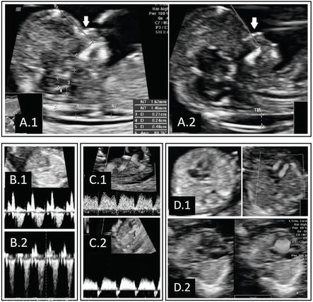
They often have leakage across the tricuspid valve and reverse flow in the ductus venosus. Dilatation of the kidneys pyelectasis.

A Review On Techniques For Computer Aided Diagnosis Of Soft Markers For Detection Of Down Syndrome In Ultrasound Fetal Images Biomedical And Pharmacology Journal
Down Syndrome can include cardiovascular central nervous craniofacial musculoskeletal gastrointestinal and urinary tract system anomalies.

What does a down syndrome ultrasound look like. What are some of the signs and ultrasound findings of down syndrome. Thickened nuchal fold nuchal translucency Duodenal Atresia double bubble Echogenic bowel. The following are ultrasound markers that are seen more frequently in fetuses with Down syndrome.
An ultrasound technician will gently move a. For most people with Down syndrome this anomaly causes a host of distinctive physical characteristics as well as potential health and medical problems. Also the lining would be thin without any masses.
Ultrasonography is the imaging modality mainstay of prenatal screening and diagnosis of Down syndrome and it is often used in combination with biochemical tests. Certain second trimester markers for Downs syndrome that are detected during an ultrasound exam are more meaningful than other markers. Ultrasound is a key component of aneuploidy screening.
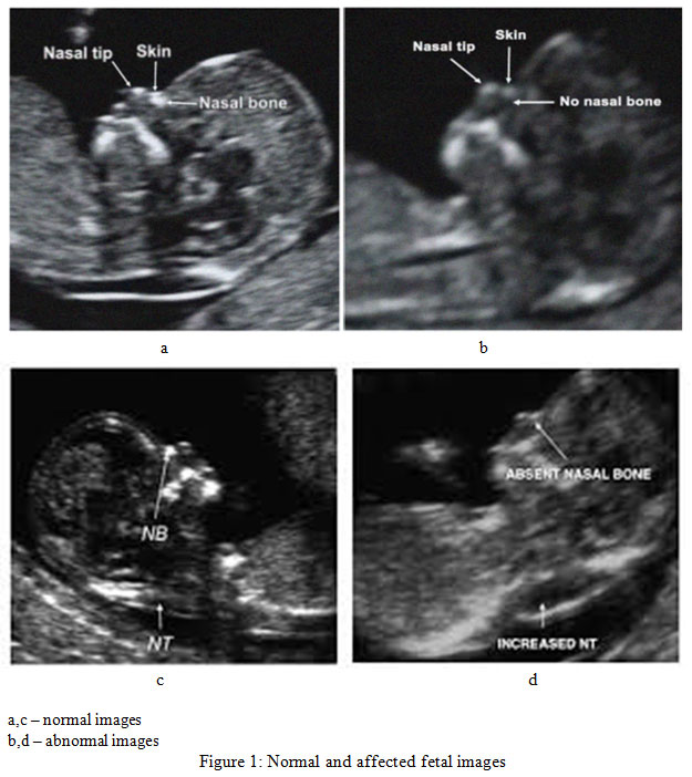
Babies with Downs syndrome are more likely to have a small or absent nose bone with a flat profile. Prenatal ultrasound findings were reviewed in 94 consecutive fetuses with proved Down syndrome trisomy 21 during a 6-year period at a single institution. The most predictive finding of Down syndrome on prenatal ultrasound is an absent nasal bone.
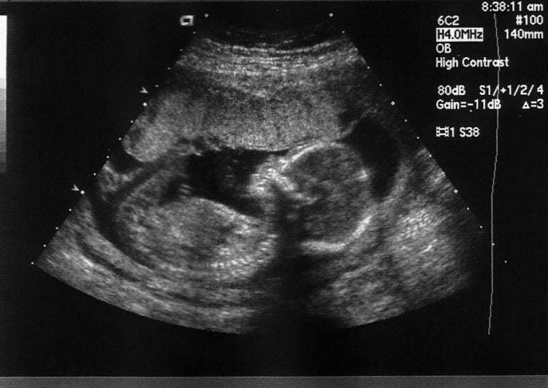
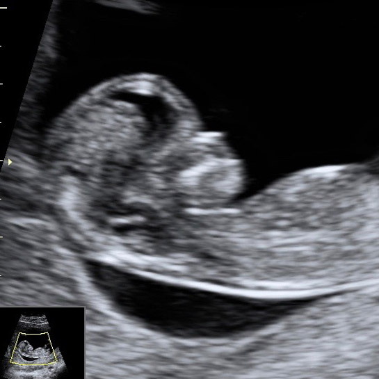
This ultrasound uses high frequency sound waves that generate a moving picture of your internal workings and your babys changing form. The nasal bone can be a good marker for Downs syndrome agrees Rosemary Reiss who practises maternal fetal medicine at Brigham and Womens Hospital in Boston Massachusetts. What does a down syndrome ultrasound look like.
The FP line was defined as the line that passes through the midpoint of the anterior border of. One or more abnormalities were found in 31 fetuses 33 including two of 11 fetuses seen before 14 weeks 17 of 68 fetuses seen between 14-24. A Verified Doctor answered.
Your age the level of two hormones free ß-hCG and PAPP-A in your blood and. The entire uterus would look the same with out any fibroid s which would look like balls. Some soft markers have a higher association with Down syndrome than others.
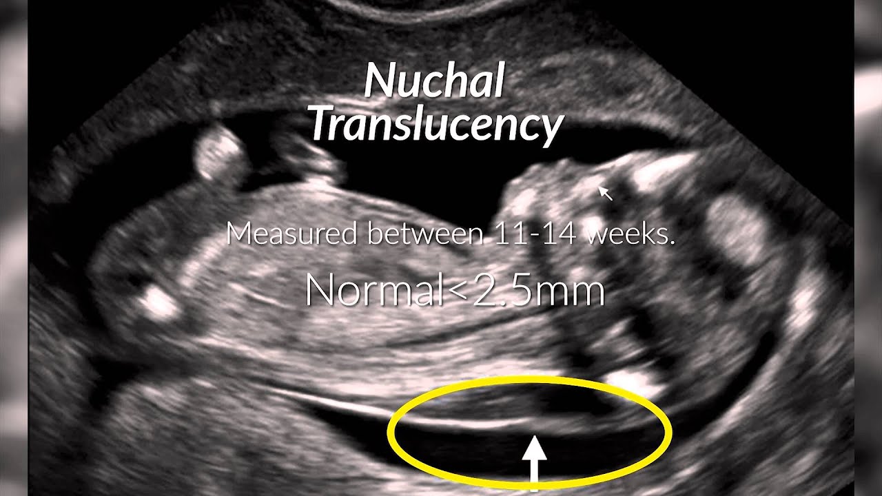
Both major structural abnormalities and minor soft markers can be detected by ultrasound in fetuses affected with aneuploidies. Between 10 and 14 weeks of your pregnancy nuchal translucency scan NTS will be performed to test the foetus for the risk of Down syndrome and other chromosomal abnormalities. Send thanks to the doctor.
In this the unborn babys nuchal fold which is the transparent back of the neck will be measured to test for any abnormalities. A 45-year-old member asked. Down syndrome is a genetic disorder in which there is an extra full or partial chromosome 21.
So adding in these additional markers will take the average detection rate of the standard technique of 80 up to 95. Especially the thickness of fluid behind the babys neck nuchal translucency thickness and possible structural anomalies. Twodimensional ultrasound images in a euploid fetus at 24 6 weeks gestation a and a fetus with Down syndrome at 28 2 weeks b showing maxillanasionmandible angle.
Second-trimester ultrasonography helps detect 60-91 cases of Down syndrome depending on. Many are spotted in fetuses without any genetic abnormalities and resolve before birth. The ultrasound examination between 11 and 13 weeks also evaluates the risk of Down syndrome which depends on.
A soft marker may indicate an increased likelihood of a chromosomal abnormality but its simply not very reliable especially considered outside of the bigger picture. A US doctor answered Learn more.

Ultrasound Scan Spots Down S Syndrome Nature News

Fetal Facial Profile Markers Of Down Syndrome In The Second And Third Trimesters Of Pregnancy Vos 2015 Ultrasound In Obstetrics Amp Gynecology Wiley Online Library

Dedicated To The Mission Of Bringing Free Or Low Cost Educational Materials And Information To The Global Ultrasound Community

Pin On Prenatal Down Syndrome Diagnosis

Soft Markers Down Syndrome April 2018 Birth Club Babycenter Canada

Finding Down Syndrome Via Ultrasound Little Doctors

Nasal Bone Length Prenasal Thickness Ratio A Strong 2d Ultrasound Marker For Down Syndrome Abstract Europe Pmc
Why Down Syndrome Is In The Middle Of The Pa Abortion Debate Whyy
Nuchal Translucency Scan 12 Week Scan Down S Syndrome Screening

Down Syndrome Screening Chromosomal Abnormality Screening Ultrasound Care

Ultrasound Images Of A Normal Fetus A And A Fetus With Trisomy 21 B Download Scientific Diagram

Ultrasound Image Classification For Down Syndrome During First Trimester Using Haralick Features Semantic Scholar

Pin On Senior Project Down Syndrome Trisomy 21

A Review Of Ultrasound Imaging Techniques For The Detection Of Down Syndrome Sciencedirect
Diagnosis Of Down Syndrome Youtube
Prenatal Diagnosis Of Down Syndrome Intechopen

5 Things To Know About Prenatal Down Syndrome Risk Tests Wsj
A Review Of Ultrasound Imaging Techniques For The Detection Of Down Syndrome Sciencedirect

Do Babies With Down S Syndrome Have No Nose Bone Babycentre Uk